| Rabbit Anti-TNFR2 antibody |
| 反应物种(预测) |
Rat |
| 产品应用(已验证) |
WB,IHC,ICC,FCM |
| 产品应用(可尝试) |
IF,IP |
| 推荐稀释比例 |
WB=1:500-2000,IP=1:10-50,IHC-P=1:50-200,IHC-F=1:50-200,Flow Cyt=1:50,IF=1:50-200,ICC=1:50, |
| 研究领域 |
肿瘤,细胞生物,神经生物学,信号转导,细胞凋亡,细胞膜受体 |
| 标签 |
Array |
-
SW480 cell; 4% Paraformaldehyde-fixed; Triton X-100 at room temperature for 20 min; Blocking buffer (normal goat serum, C-0005) at 37°C for 20 min; Antibody incubation with (TNF Receptor II) monoclonal Antibody, Unconjugated (bsm-52938R) 1:50, 90 minutes at 37°C; followed by a conjugated Goat Anti-Rabbit IgG antibody at 37°C for 90 minutes, DAPI (blue, C02-04002) was used to stain the cell nuclei.
-
Blank control:HL-60.
Primary Antibody (green line): Rabbit Anti-TNF Receptor II antibody (bsm-52938R)
Dilution: 1:50;
Isotype Control Antibody (orange line): Rabbit IgG .
Secondary Antibody : Goat anti-rabbit IgG-AF488
Dilution: 1:1000.
Protocol
The cells were fixed with 4% PFA (10min at room temperature)and then permeabilized with 0.1% PBST for 20 min at room temperature. The cells were then incubated in 5%BSA to block non-specific protein-protein interactions for 30 min at room temperature .Cells stained with Primary Antibody for 30 min at room temperature. The secondary antibody used for 40 min at room temperature. Acquisition of 20,000 events was performed.
-
Sample:
Lane 1: MCF-7 cell lysate
Lane 2: Jurkat cell lysate
Lane 3: Hela cell lysate
Primary: Anti-TNFR2 (bsm-52938R) at 1:500 dilution
Secondary: Goat Anti-Rabbit IgG - HRP at 1:5000 dilution
Predicted band size: 46 kD
Observed band size: 60 kD
-
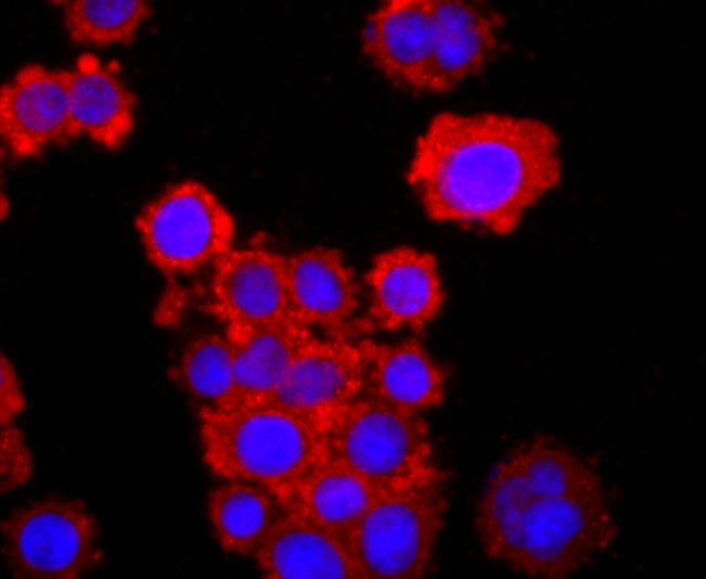
MCF-7 cell; 4% Paraformaldehyde-fixed; Triton X-100 at room temperature for 20 min; Blocking buffer (normal goat serum, C-0005) at 37°C for 20 min; Antibody incubation with (TNF Receptor II) monoclonal Antibody, Unconjugated (bsm-52938R) 1:50, 90 minutes at 37°C; followed by a conjugated Goat Anti-Rabbit IgG antibody at 37°C for 90 minutes, DAPI (blue, C02-04002) was used to stain the cell nuclei.
-
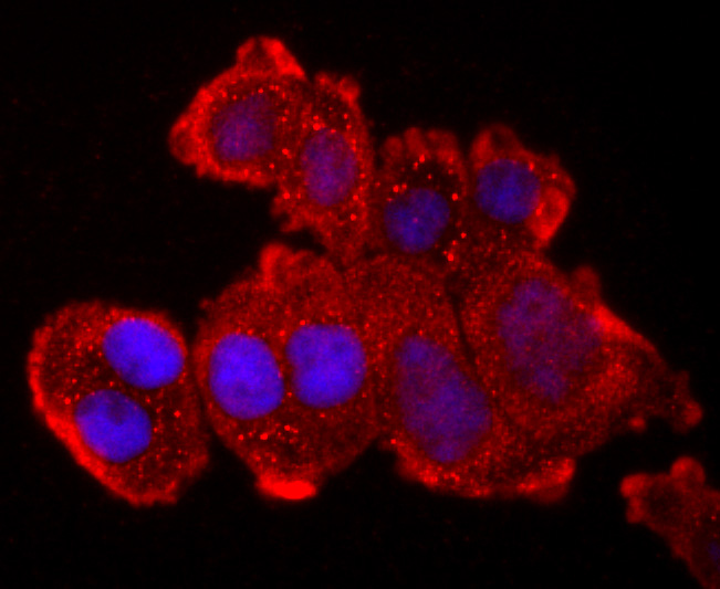
Hela cell; 4% Paraformaldehyde-fixed; Triton X-100 at room temperature for 20 min; Blocking buffer (normal goat serum, C-0005) at 37°C for 20 min; Antibody incubation with (TNF Receptor II) monoclonal Antibody, Unconjugated (bsm-52938R) 1:50, 90 minutes at 37°C; followed by a conjugated Goat Anti-Rabbit IgG antibody at 37°C for 90 minutes, DAPI (blue, C02-04002) was used to stain the cell nuclei.
-
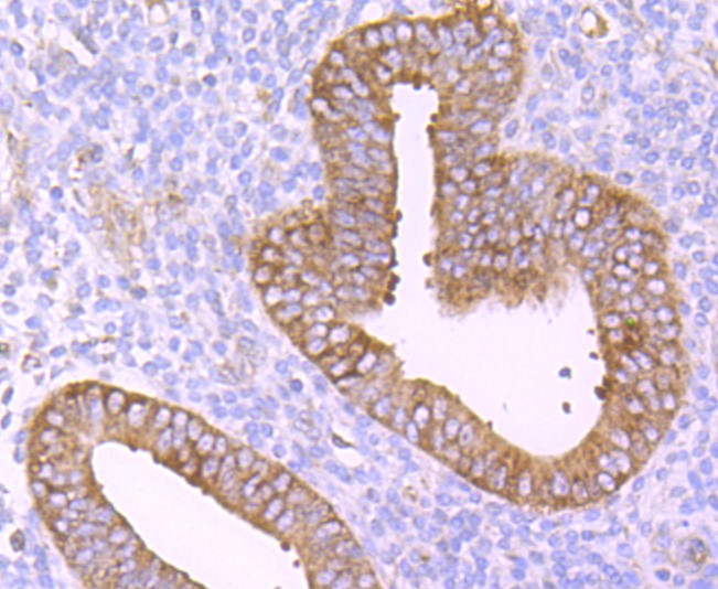
Paraformaldehyde-fixed, paraffin embedded (human uterus tissue); Antigen retrieval by boiling in sodium citrate buffer (pH6.0) for 15min; Block endogenous peroxidase by 3% hydrogen peroxide for 20 minutes; Blocking buffer (normal goat serum) at 37°C for 30min; Antibody incubation with (TNFR2) Monoclonal Antibody, Unconjugated (bsm-52938R) at 1:50 overnight at 4°C, followed by operating according to SP Kit(Rabbit) (sp-0023) instructionsand DAB staining.
-
Paraformaldehyde-fixed, paraffin embedded (mouse kidney tissue); Antigen retrieval by boiling in sodium citrate buffer (pH6.0) for 15min; Block endogenous peroxidase by 3% hydrogen peroxide for 20 minutes; Blocking buffer (normal goat serum) at 37°C for 30min; Antibody incubation with (TNFR2) Monoclonal Antibody, Unconjugated (bsm-52938R) at 1:50 overnight at 4°C, followed by operating according to SP Kit(Rabbit) (sp-0023) instructionsand DAB staining.
-
Paraformaldehyde-fixed, paraffin embedded (human kidney tissue); Antigen retrieval by boiling in sodium citrate buffer (pH6.0) for 15min; Block endogenous peroxidase by 3% hydrogen peroxide for 20 minutes; Blocking buffer (normal goat serum) at 37°C for 30min; Antibody incubation with (TNFR2) Monoclonal Antibody, Unconjugated (bsm-52938R) at 1:50 overnight at 4°C, followed by operating according to SP Kit(Rabbit) (sp-0023) instructionsand DAB staining.
-
Sample:
Lane 1: Human THP-1 cell Lysates
Lane 2: Human K562 cell Lysates
Lane 3: Human U937 cell Lysates
Lane 4: Human U87MG cell Lysates
Lane 5: Human HUVEC cell Lysates
Primary: Anti-TNFR2 (bsm-52938R) at 1/1000 dilution
Secondary: IRDye800CW Goat Anti-Rabbit IgG at 1/20000 dilution
Predicted band size: 46kDa
Observed band size: 70 kDa
RRID:RRID
产品名称:Rabbit Anti-TNFR2 antibody
别名: TNF Receptor II; CD120b; p75; p75TNFR; p80 TNF alpha receptor; p80 TNF-alpha receptor; TBP-2; TBPII; TNF R2; TNF R75; TNF-R2; TNF-RII; TNFR-II; TNFR80; TNFRII; TNFRSF1B; TNR1B_HUMAN; Tumor necrosis factor receptor 2; Tumor necrosis factor receptor type II
中文名称:肿瘤坏死因子受体2重组兔单克隆抗体
英文名称:Rabbit Anti-TNFR2 antibody
抗体来源: Rabbit
克隆类型:单克隆
细胞定位:细胞膜,分泌型蛋白
性 状:Liquid
亚 型:IgG
纯化方法:affinity purified by Protein A
保存条件:Shipped at 4℃. Store at -20 °C for one year. Avoid repeated freeze/thaw cycles.
免 疫 原:KLH conjugated synthetic peptide derived from human TNFR2
SWISS:P20333
Gene ID :7133
Human Gene ID:7133
The protein encoded by this gene is a member of the TNF-receptor superfamily. This protein and TNF-receptor 1 form a heterocomplex that mediates the recruitment of two anti-apoptotic proteins, c-IAP1 and c-IAP2, which possess E3 ubiquitin ligase activity. The function of IAPs in TNF-receptor signalling is unknown, however, c-IAP1 is thought to potentiate TNF-induced apoptosis by the ubiquitination and degradation of TNF-receptor-associated factor 2, which mediates anti-apoptotic signals. Knockout studies in mice also suggest a role of this protein in protecting neurons from apoptosis by stimulating antioxidative pathways. [provided by RefSeq, Jul 2008]
Function:Receptor with high affinity for TNFSF2/TNF-alpha and approximately 5-fold lower affinity for homotrimeric TNFSF1/lymphotoxin-alpha. The TRAF1/TRAF2 complex recruits the apoptotic suppressors BIRC2 and BIRC3 to TNFRSF1B/TNFR2. This receptor mediates most o
Subcellular Location:Secreted and Cell membrane.
Post-translational modifications:Phosphorylated; mainly on serine residues and with a very low level on threonine residues.
A soluble form (tumor necrosis factor binding protein 2) is produced from the membrane form by proteolytic processing.
Similarity:Contains 4 TNFR-Cys repeats.
Important Note:This product as supplied is intended for research use only, not for use in human, therapeutic or diagnostic applications.
 400-901-9800


 说明书
说明书 联系我们
联系我们 打印此页面
打印此页面 收藏
收藏