| Rabbit Anti-MCL1 antibody |
| 反应物种(预测) |
Dog,Pig,Horse,Rabbit |
| 产品应用(已验证) |
WB,IHC,FCM |
| 产品应用(可尝试) |
ICC,IF |
| 推荐稀释比例 |
WB=1:500-2000,IHC-P=1:100-500,IHC-F=1:100-500,Flow Cyt=1ug/Test,IF=1:100-500,ICC=1:100-500, |
| 研究领域 |
肿瘤,免疫学,信号转导,细胞凋亡,转录调节因子,线抗体, |
| 标签 |
Array |
-
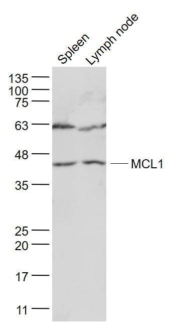
Sample:
Spleen (Mouse) Lysate at 40 ug
Lymph node (Mouse) Lysate at 40 ug
Primary: Anti- MCL1 (bs-23315R) at 1/1000 dilution
Secondary: IRDye800CW Goat Anti-Rabbit IgG at 1/20000 dilution
Predicted band size: 39 kD
Observed band size: 39 kD
-
Sample:
Lane 1: Lymph node (Mouse) Lysate at 40 ug
Lane 2: Spleen (Mouse) Lysate at 40 ug
Lane 3: Small intestine (Mouse) Lysate at 40 ug
Lane 4: NIH/3T3 (Mouse) Cell Lysate at 30 ug
Lane 5: Lymph node (Rat) Lysate at 40 ug
Lane 6: Spleen (Rat) Lysate at 40 ug
Lane 7: Small intestine (Rat) Lysate at 40 ug
Lane 8: Bone (Rat) Lysate at 40 ug
Lane 9: HL60 (Human) Cell Lysate at 30 ug
Lane 10: A431 (Human) Cell Lysate at 30 ug
Lane 11: K562 (Human) Cell Lysate at 30 ug
Lane 12: MDA-MB-231 (Human) Cell Lysate at 30 ug
Lane 13: Raji (Human) Cell Lysate at 30 ug
Lane 14: A549 (Human) Cell Lysate at 30 ug
Primary: Anti-MCL1 (bs-23315R) at 1/1000 dilution
Secondary: IRDye800CW Goat Anti-Rabbit IgG at 1/20000 dilution
Predicted band size: 39 kD
Observed band size: 40 kD
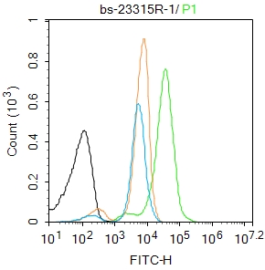
-
Blank control: K562.
Primary Antibody (green line): Rabbit Anti-MCL1 antibody (bs-23315R)
Dilution: 1μg /10^6 cells;
Isotype Control Antibody (orange line): Rabbit IgG .
Secondary Antibody : Goat anti-rabbit IgG-FITC
Dilution: 1μg /test.
Protocol
The cells were fixed with 4% PFA (10min at room temperature)and then permeabilized with 90% ice-cold methanol for 20 min at-20℃. The cells were then incubated in 5%BSA to block non-specific protein-protein interactions for 30 min at room temperature .Cells stained with Primary Antibody for 30 min at room temperature. The secondary antibody used for 40 min at room temperature. Acquisition of 20,000 events was performed.
-
Sample:
NIH/3T3(Mouse) Cell Lysate at 30 ug
Primary: Anti-MCL1 (bs-23315R) at 1/1000 dilution
Secondary: IRDye800CW Goat Anti-Rabbit IgG at 1/20000 dilution
Predicted band size: 39 kD
Observed band size: 41 kD
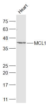
-
Sample:
Heart (Rat) Lysate at 40 ug
Primary: Anti-MCL1 (bs-23315R) at 1/1000 dilution
Secondary: IRDye800CW Goat Anti-Rabbit IgG at 1/20000 dilution
Predicted band size: 39 kD
Observed band size: 41 kD
-
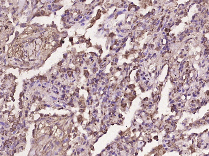
Paraformaldehyde-fixed, paraffin embedded (human lung carcinoma); Antigen retrieval by boiling in sodium citrate buffer (pH6.0) for 15min; Block endogenous peroxidase by 3% hydrogen peroxide for 20 minutes; Blocking buffer (normal goat serum) at 37°C for 30min; Antibody incubation with (MCL1) Polyclonal Antibody, Unconjugated (bs-23315R) at 1:400 overnight at 4°C, followed by operating according to SP Kit(Rabbit) (sp-0023) instructionsand DAB staining.
-
Paraformaldehyde-fixed, paraffin embedded (Mouse embryo); Antigen retrieval by boiling in sodium citrate buffer (pH6.0) for 15min; Block endogenous peroxidase by 3% hydrogen peroxide for 20 minutes; Blocking buffer (normal goat serum) at 37°C for 30min; Antibody incubation with (MCL1) Polyclonal Antibody, Unconjugated (bs-23315R) at 1:400 overnight at 4°C, followed by operating according to SP Kit(Rabbit) (sp-0023) instructions and DAB staining.
-
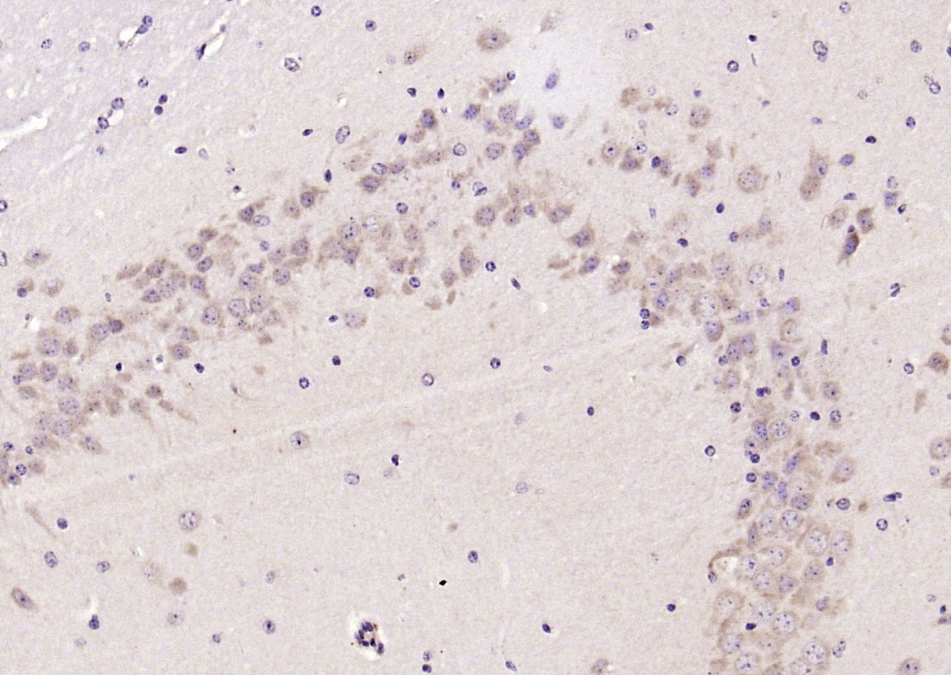
Paraformaldehyde-fixed, paraffin embedded (mouse brain); Antigen retrieval by boiling in sodium citrate buffer (pH6.0) for 15min; Block endogenous peroxidase by 3% hydrogen peroxide for 20 minutes; Blocking buffer (normal goat serum) at 37°C for 30min; Antibody incubation with (MCL1) Polyclonal Antibody, Unconjugated (bs-23315R) at 1:200 overnight at 4°C, followed by operating according to SP Kit(Rabbit) (sp-0023) instructionsand DAB staining.
-
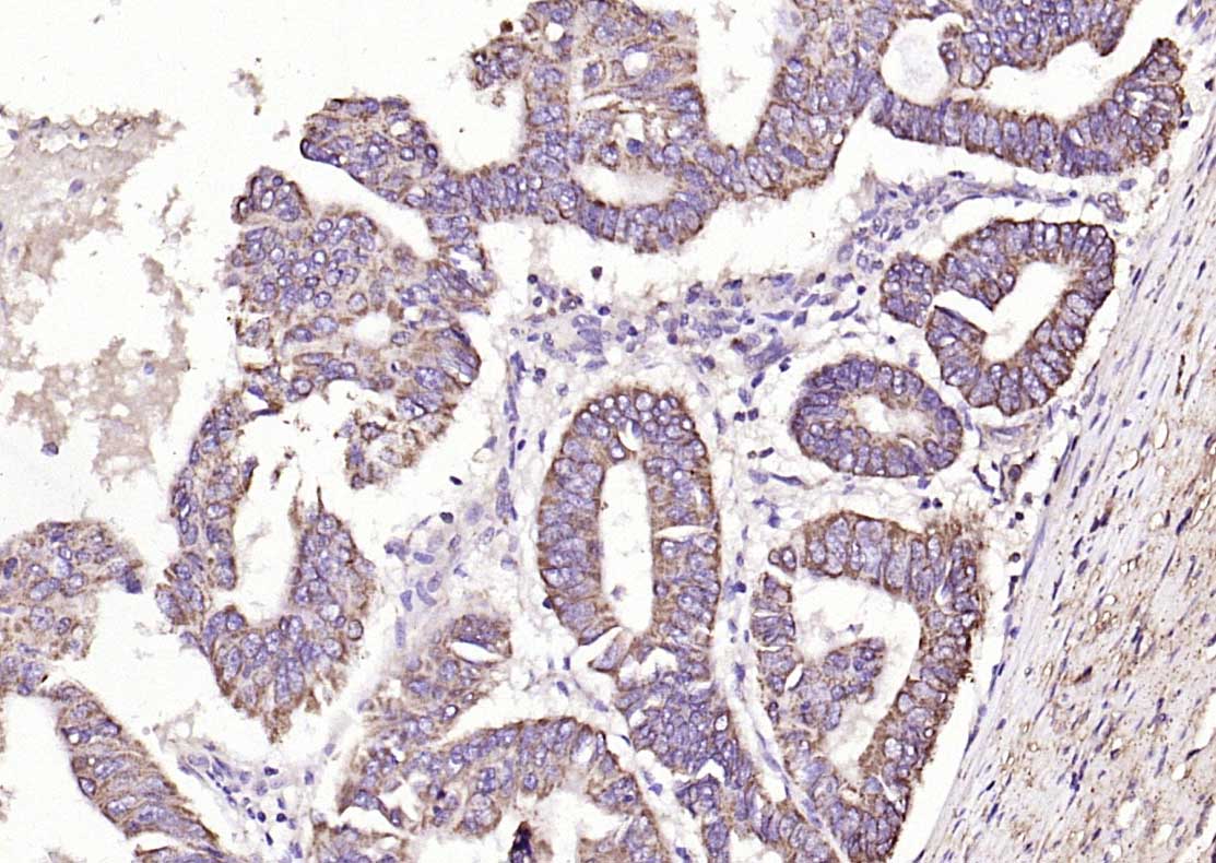
Paraformaldehyde-fixed, paraffin embedded (human colon carcinoma); Antigen retrieval by boiling in sodium citrate buffer (pH6.0) for 15min; Block endogenous peroxidase by 3% hydrogen peroxide for 20 minutes; Blocking buffer (normal goat serum) at 37°C for 30min; Antibody incubation with (MCL1) Polyclonal Antibody, Unconjugated (bs-23315R) at 1:200 overnight at 4°C, followed by operating according to SP Kit(Rabbit) (sp-0023) instructionsand DAB staining.
RRID:RRID
产品名称:Rabbit Anti-MCL1 antibody
别名: myeloid cell leukemia 1; myeloid cell leukemia sequence 1; MCL-1; MCL1L; MCL 1; mcl1/EAT; MGC104264; MGC1839; TM; MCL1S; EAT) Bcl 2 related protein EAT/mcl1; BCL2 related; BCL2L3; EAT; Induced myeloid leukemia cell differentiation protein Mcl 1; myeloid c
中文名称:髓样细胞白血病-1抗体
英文名称:Rabbit Anti-MCL1 antibody
抗体来源: Rabbit
克隆类型:多克隆
细胞定位:细胞核,细胞浆,细胞膜
性 状:Liquid
亚 型:IgG
纯化方法:affinity purified by Protein A
保存条件:Shipped at 4℃. Store at -20 °C for one year. Avoid repeated freeze/thaw cycles.
免 疫 原:KLH conjugated synthetic peptide derived from human MCL1
抗原表位:101-200/350
SWISS:Q07820
Gene ID :4170
Human Gene ID:4170
Mcl1 is an anti-apoptotic member of Bcl2 family originally isolated from the ML1 human myeloid leukemia cell line during phorbol ester-induced differentiation along the monocyte/macrophage pathway. Mcl1 localizes to the mitochondria, interacts with and antagonizes pro-apoptotic Bcl2 family members, and inhibits apoptosis by a number of cytotoxic stimuli. It is involved in programing of differentiation and concomitant maintenance of viability but not of proliferation. Isoform 1 inhibits apoptosis while isoform 2 promotes it. Expression increases early during phorbol-ester induced differentiation along the monocyte/macrophage pathway in myeloid leukemia cell lines ML1.
Function:Involved in the regulation of apoptosis versus cell survival, and in the maintenance of viability but not of proliferation. Mediates its effects by interactions with a number of other regulators of apoptosis. Isoform 1 inhibits apoptosis. Isoform 2 promot
Subunit:Interacts with BAD, BOK, BIK and BFM (By similarity). Interacts with PMAIP1. Isoform 1 interacts with BAX, BAK1, TPT1 and BCL2L11. Heterodimer of isoform 1 and isoform 2. Homodimers of isoform 1 or isoform 2 are not detected. Isoform 2 does not interact w
Subcellular Location:Membrane; Single-pass membrane protein (Potential). Cytoplasm. Mitochondrion. Nucleus, nucleoplasm. Note=Cytoplasmic, associated with mitochondria.
Post-translational modifications:Cleaved by CASP3 during apoptosis. In intact cells cleavage occurs preferentially after Asp-127, yielding a pro-apoptotic 28 kDa C-terminal fragment.
Rapidly degraded in the absence of phosphorylation on Thr-163 in the PEST region.
Phosphorylate
Similarity:Belongs to the Bcl-2 family.
Important Note:This product as supplied is intended for research use only, not for use in human, therapeutic or diagnostic applications.
 400-901-9800


 说明书
说明书 联系我们
联系我们 打印此页面
打印此页面 收藏
收藏