| Rabbit Anti-Cytokeratin 13 antibody |
| 反应物种(预测) |
Rat,Dog,Horse,Rabbit,Sheep |
| 产品应用(已验证) |
WB |
| 产品应用(可尝试) |
ELISA |
| 推荐稀释比例 |
WB=1:500-2000,Elisa=1:5000-10000, |
| 研究领域 |
肿瘤 |
| 标签 |
Array |
-
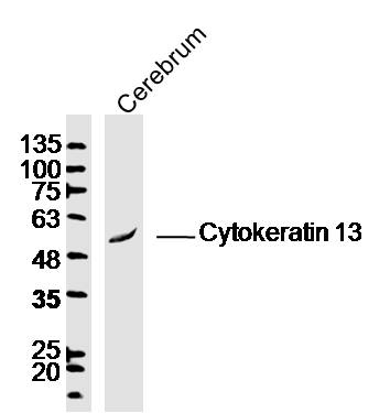
Sample:Cerebrum (Mouse)Lysate at 40 ug
Primary: Anti-Cytokeratin 13(bs-1717R)at 1/300 dilution
Secondary: IRDye800CW Goat Anti-RabbitIgG at 1/20000 dilution
Predicted band size: 49kD
Observed band size: 49kD
-
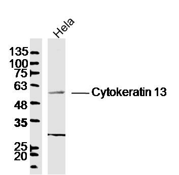
Sample: Hela (Human) Cell Lysate at 40 ug
Primary: Anti-Cytokeratin 13(bs-1717R)at 1/300 dilution
Secondary: IRDye800CW Goat Anti-RabbitIgG at 1/20000 dilution
Predicted band size: 49kD
Observed band size: 55kD
-
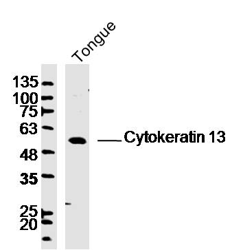
Sample: Tongue(Mouse)Lysate at 40 ug
Primary: Anti-Cytokeratin 13(bs-1717R)at 1/300 dilution
Secondary: IRDye800CW Goat Anti-RabbitIgG at 1/20000 dilution
Predicted band size: 49kD
Observed band size: 55kD
RRID:AB_10855891
产品名称:Rabbit Anti-Cytokeratin 13 antibody
别名: type I cytoskeletal 13; 47 kDa cytokeratin; CK-13; CK13; Cytokeratin-13; K13; K1C13_HUMAN; Ka13; Keratin 13; Keratin; keratin type I cytoskeletal 13; Keratin-13; Krt-1.13; Krt1-13; KRT13; MGC161462; MGC3781; Type I keratin Ka13; Keratin, type I cytoskelet
中文名称:细胞角蛋白13抗体
英文名称:Rabbit Anti-Cytokeratin 13 antibody
抗体来源: Rabbit
克隆类型:多克隆
细胞定位:细胞浆
性 状:Liquid
亚 型:IgG
纯化方法:affinity purified by Protein A
保存条件:Shipped at 4℃. Store at -20 °C for one year. Avoid repeated freeze/thaw cycles.
免 疫 原:KLH conjugated synthetic peptide derived from human Cytokeratin-13
抗原表位:251-350/458
SWISS:P13646
Gene ID :3860
Human Gene ID:3860
The protein encoded by this gene is a member of the keratin gene family. The keratins are intermediate filament proteins responsible for the structural integrity of epithelial cells and are subdivided into cytokeratins and hair keratins. Most of the type I cytokeratins consist of acidic proteins which are arranged in pairs of heterotypic keratin chains. This type I cytokeratin is paired with keratin 4 and expressed in the suprabasal layers of non-cornified stratified epithelia. Mutations in this gene and keratin 4 have been associated with the autosomal dominant disorder White Sponge Nevus. The type I cytokeratins are clustered in a region of chromosome 17q21.2. Alternative splicing of this gene results in multiple transcript variants; however, not all variants have been described. [provided by RefSeq, Jul 2008].
Subunit:Heterotetramer of two type I and two type II keratins. keratin-13 is generally associated with keratin-4.
Tissue Specificity:Defects in KRT13 are a cause of white sponge nevus of cannon (WSN) . WSN is a rare autosomal dominant disorder which predominantly affects non-cornified stratified squamous epithelia. Clinically, it is characterized by the presence of soft, white, and spo
DISEASE:White sponge nevus of cannon (WSN) [MIM:193900]: Rare autosomal dominant disorder which predominantly affects non-cornified stratified squamous epithelia. Clinically, it is characterized by the presence of soft, white, and spongy plaques in the oral mucos
Similarity:Belongs to the intermediate filament family.
Important Note:This product as supplied is intended for research use only, not for use in human, therapeutic or diagnostic applications.
 400-901-9800


 说明书
说明书 联系我们
联系我们 打印此页面
打印此页面 收藏
收藏