| Rabbit Anti-HAS1 antibody |
| 反应物种(预测) |
Dog,Pig,Cow |
| 产品应用(已验证) |
WB,FCM |
| 产品应用(可尝试) |
IHC,ICC,IF,ELISA |
| 推荐稀释比例 |
WB=1:500-2000,Elisa=1:5000-10000,IHC-P=1:100-500,IHC-F=1:100-500,Flow Cyt=1ug/Test,IF=1:100-500,ICC=1:100-500, |
| 研究领域 |
心血管,细胞生物,发育生物学,神经生物学,信号转导,干细胞,细胞粘附分子, |
| 标签 |
Array |
-
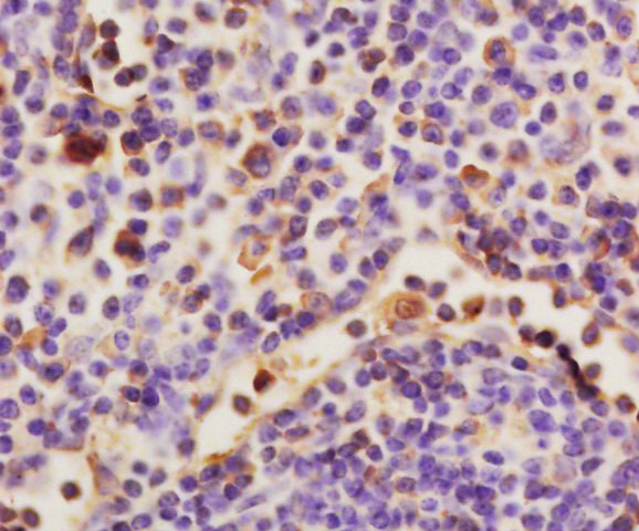
Tissue/cell: rat spleen tissue; 4% Paraformaldehyde-fixed and paraffin-embedded;
Antigen retrieval: citrate buffer ( 0.01M, pH 6.0 ), Boiling bathing for 15min; Block endogenous peroxidase by 3% Hydrogen peroxide for 30min; Blocking buffer (normal goat serum,C-0005) at 37℃ for 20 min;
Incubation: Anti-HAS1 Polyclonal Antibody, Unconjugated(bs-2946R) 1:200, overnight at 4°C, followed by conjugation to the secondary antibody(SP-0023) and DAB(C-0010) staining
-
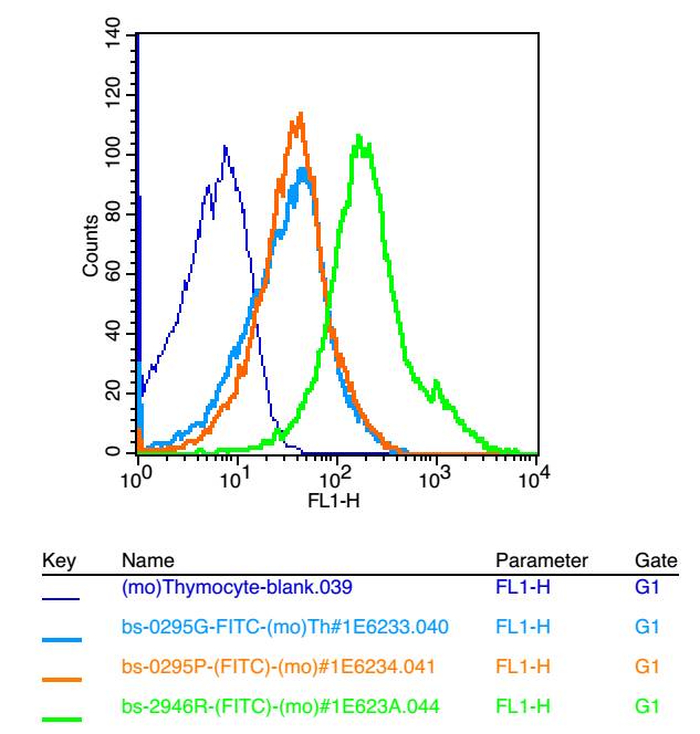
Blank control: mouse thymouses(blue)
Isotype Control Antibody: Rabbit IgG(orange) ;
Secondary Antibody: Goat anti-rabbit IgG-FITC(white blue),
Dilution: 1:100 in 1 X PBS containing 0.5% BSA ;
Primary Antibody Dilution: 1μl in 100 μL1X PBS containing 0.5% BSA(green).
-
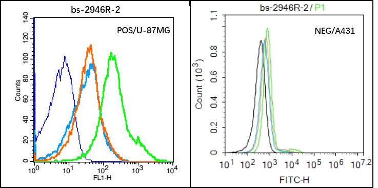
Black line : Positive blank control (U87MG); Negative blank control (A431)
Green line : Primary Antibody (Rabbit Anti-HAS1 antibody (bs-2946R) )
Orange line:Isotype Control Antibody (Rabbit IgG) .
Blue line : Secondary Antibody (Goat anti-rabbit IgG-AF488)
U87MG(Positive)and A431(Negative control)cells (black) were incubated in 5% BSA blocking buffer for 30 min at room temperature. Cells were then stained with HAS1 Antibody(bs-2946R)at 1:50 dilution in blocking buffer and incubated for 30 min at room temperature, washed twice with 2% BSA in PBS, followed by secondary antibody(blue) incubation for 40 min at room temperature. Acquisitions of 20,000 events were performed. Cells stained with primary antibody (green), and isotype control (orange).
-
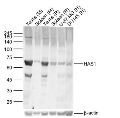
Sample:
Lane 1: Mouse Testis Lysates
Lane 2: Mouse Spleen Lysates
Lane 3: Rat Testis Lysates
Lane 4: Rat Spleen Lysates
Lane 5: Human U-87 MG cell Lysates
Lane 6: Human DU145 cell Lysates
Primary: Anti-HAS1 (bs-2946R) at 1/1000 dilution
Secondary: IRDye800CW Goat Anti-Rabbit IgG at 1/20000 dilution
Predicted band size: 65kDa
Observed band size: 65kDa
RRID:AB_11081524
产品名称:Rabbit Anti-HAS1 antibody
别名: HA synthase 1; HAS; HAS1; HAS 1; HAS-1; HAS1_HUMAN; HuHAS1; Hyaluronan synthase 1; Hyaluronan synthase; HA Synthase: Hyaluronate synthase 1; Hyaluronic acid synthase 1.
中文名称:透明质酸合成酶1抗体
英文名称:Rabbit Anti-HAS1 antibody
抗体来源: Rabbit
克隆类型:多克隆
细胞定位:细胞膜
性 状:Liquid
亚 型:IgG
纯化方法:affinity purified by Protein A
保存条件:Shipped at 4℃. Store at -20 °C for one year. Avoid repeated freeze/thaw cycles.
免 疫 原:KLH conjugated synthetic peptide derived from human Hyaluronan synthase 1
抗原表位:501-578/578
抗原细胞定位:Extracellular
SWISS:Q92839
Gene ID :3036
Human Gene ID:3036
HAS1, HAS2 and HAS3 are HA Synthase proteins that synthesize HA (Hyaluronan or hyaluronic acid). The extracellular matrix in most vertebrates express HA, which is a high molecular weight linear polysaccharide composed of alternating glucuronic acid and N-acetylglucosamine residues linked by beta-1,3 and beta-1,4 glycosidic bonds. The three HAS genes show distinct patterns of expression during development and their protein products play significantly different roles in the formation of the HA matrix. Both HAS1 and HAS2 synthesize high molecular weight HA, whereas HAS3 produces lower molecular weight HA. The expression of the three HAS isoforms is more prominent in growing cells than in resting cells and is differentially regulated by various stimuli, suggesting distinct functional roles of the three proteins. HAS1 mRNA shows predominant expression in bone marrow mesenchymal progenitor cells and synovial cells.
Function:Plays a role in hyaluronan/hyaluronic acid (HA) synthesis. Also able to catalyze the synthesis of chito-oligosaccharide depending on the substrate.
Subcellular Location:Membrane; Multi-pass membrane protein
Tissue Specificity:Highly expressed in ovary followed by spleen, thymus, prostate, testes and large intestine. Weakly expressed in small intestine.
Similarity:Belongs to the NodC/HAS family.
Important Note:This product as supplied is intended for research use only, not for use in human, therapeutic or diagnostic applications.
 400-901-9800


 说明书
说明书 联系我们
联系我们 打印此页面
打印此页面 收藏
收藏