| Rabbit Anti-MAP1A antibody |
| 反应物种(预测) |
Mouse,Chicken,Dog,Pig,Cow,Horse |
| 产品应用(已验证) |
IHC,FCM |
| 产品应用(可尝试) |
IF |
| 推荐稀释比例 |
IHC-P=1:100-500,IHC-F=1:100-500,Flow Cyt=1μg /test,IF=1:100-500, |
| 研究领域 |
肿瘤,细胞生物,免疫学,神经生物学,信号转导,细胞凋亡,激酶和磷酸酶 |
| 标签 |
Array |
-
Paraformaldehyde-fixed, paraffin embedded (Rat liver); Antigen retrieval by boiling in sodium citrate buffer (pH6.0) for 15min; Block endogenous peroxidase by 3% hydrogen peroxide for 20 minutes; Blocking buffer (normal goat serum) at 37°C for 30min; Antibody incubation with (MAP1A) Polyclonal Antibody, Unconjugated (bs-1847R) at 1:400 overnight at 4°C, followed by operating according to SP Kit(Rabbit) (sp-0023) instructionsand DAB staining.
-
Blank control (blue line): U251 (blue).
Primary Antibody (green line): Rabbit Anti- MAP1A antibody (bs-1847R)
Dilution: 1μg /10^6 cells;
Isotype Control Antibody (orange line): Rabbit IgG .
Secondary Antibody (white blue line): Goat anti-rabbit IgG-PE
Dilution: 1μg /test.
Protocol
The cells were fixed with 2% paraformaldehyde (10 min , then permeabilized) with 90% ice-cold methanol for 20 min on ice. Cells stained with Primary Antibody for 30 min at room temperature. The cells were then incubated in 1 X PBS/2%BSA/10% goat serum to block non-specific protein-protein interactions followed by the antibody for 15 min at room temperature. The secondary antibody used for 40 min at room temperature. Acquisition of 20,000 events was performed.
-
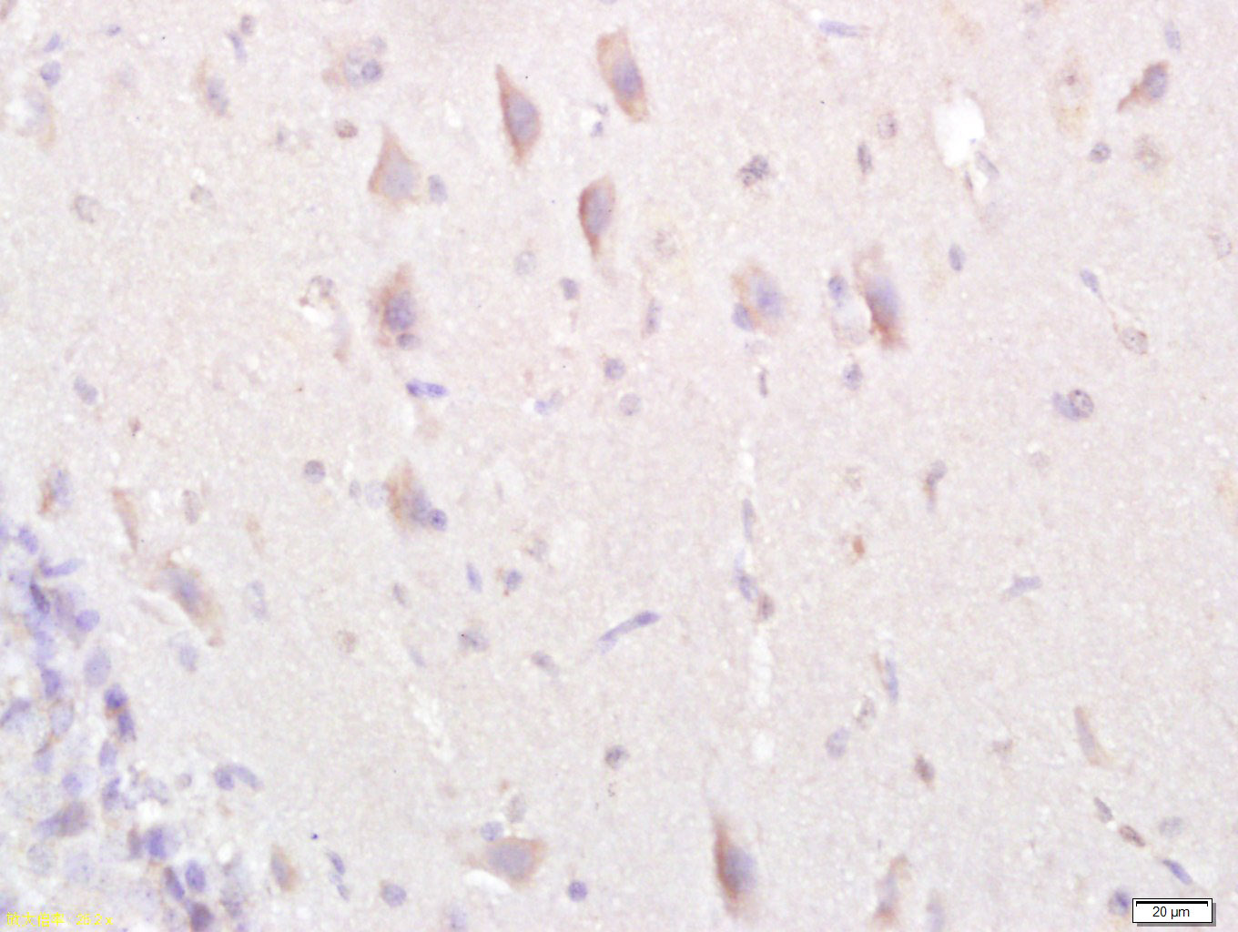
Tissue/cell: rat brain tissue; 4% Paraformaldehyde-fixed and paraffin-embedded;
Antigen retrieval: citrate buffer ( 0.01M, pH 6.0 ), Boiling bathing for 15min; Block endogenous peroxidase by 3% Hydrogen peroxide for 30min; Blocking buffer (normal goat serum,C-0005) at 37℃ for 20 min;
Incubation: Anti-MAP1A Polyclonal Antibody, Unconjugated(bs-1847R) 1:500, overnight at 4°C, followed by conjugation to the secondary antibody(SP-0023) and DAB(C-0010) staining
-
Tissue/cell: human cervical carcinoma; 4% Paraformaldehyde-fixed and paraffin-embedded;
Antigen retrieval: citrate buffer ( 0.01M, pH 6.0 ), Boiling bathing for 15min; Block endogenous peroxidase by 3% Hydrogen peroxide for 30min; Blocking buffer (normal goat serum,C-0005) at 37℃ for 20 min;
Incubation: Anti-MAP1A Polyclonal Antibody, Unconjugated(bs-1847R) 1:500, overnight at 4°C, followed by conjugation to the secondary antibody(SP-0023) and DAB(C-0010) staining
RRID:AB_10855163
产品名称:Rabbit Anti-MAP1A antibody
别名: MAP 1A; MAP-1A; MTAP1A; MAP 1L; MAP1L; MAP1A heavy chain; Map1a; MAP1A_HUMAN; Microtubule associated protein 1 like; Microtubule Associated Protein 1A; Microtubule-associated protein 1A; MTAP1A; Proliferation related protein p80; Proliferation-related pro
中文名称:微管相关蛋白1A抗体
英文名称:Rabbit Anti-MAP1A antibody
抗体来源: Rabbit
克隆类型:多克隆
细胞定位:细胞浆
性 状:Liquid
亚 型:IgG
纯化方法:affinity purified by Protein A
保存条件:Shipped at 4℃. Store at -20 °C for one year. Avoid repeated freeze/thaw cycles.
免 疫 原:KLH conjugated synthetic peptide derived from human MAP1A heavy chain
抗原表位:2651-2750/3014
SWISS:P78559
Gene ID :4130
Human Gene ID:4130
This gene encodes a protein that belongs to the microtubule-associated protein family. The proteins of this family are thought to be involved in microtubule assembly, which is an essential step in neurogenesis. The product of this gene is a precursor polypeptide that presumably undergoes proteolytic processing to generate the final MAP1A heavy chain and LC2 light chain. Expression of this gene is almost exclusively in the brain. Studies of the rat microtubule-associated protein 1A gene suggested a role in early events of spinal cord development. [provided by RefSeq, Jul 2008]
Function:Structural protein involved in the filamentous cross-bridging between microtubules and other skeletal elements.
Subunit:3 different light chains, LC1, LC2 and LC3, can associate with MAP1A and MAP1B proteins. Interacts with TIAM2. Interacts with guanylate kinase-like domain of DLG1, DLG2, DLG4. Binds to CSNK1D.
Subcellular Location:Cytoplasm, cytoskeleton (Probable).
Tissue Specificity:Brain
Post-translational modifications:Phosphorylated by CSNK1D.
LC2 is generated from MAP1A by proteolytic processing.
Similarity:Belongs to the MAP1 family.
Important Note:This product as supplied is intended for research use only, not for use in human, therapeutic or diagnostic applications.
 400-901-9800


 说明书
说明书 联系我们
联系我们 打印此页面
打印此页面 收藏
收藏