| Rabbit Anti-Cytokeratin 4 antibody |
| 反应物种(预测) |
Mouse,Dog,Cow,Horse,Rabbit |
| 产品应用(已验证) |
WB,IHC,FCM |
| 产品应用(可尝试) |
IF,ELISA |
| 推荐稀释比例 |
WB=1:500-2000,Elisa=1:5000-10000,IHC-P=1:100-500,IHC-F=1:100-500,Flow Cyt=1ug/test,IF=1:100-500, |
| 研究领域 |
肿瘤,信号转导,上皮细胞, |
| 标签 |
Array |
-
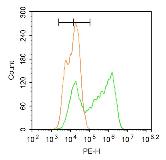
Blank control:A549.
Primary Antibody (green line): Rabbit Anti-Cytokeratin 4, membrane-bound isoform antibody (bs-1006R)
Dilution: 1μg /10^6 cells;
Isotype Control Antibody (orange line): Rabbit IgG .
Secondary Antibody : Goat anti-rabbit IgG-PE
Dilution: 3μg /test.
Protocol
The cells were fixed with 4% PFA (10min at room temperature)and then permeabilized with 90% ice-cold methanol for 20 min at-20℃. The cells were then incubated in 5% BSA to block non-specific protein-protein interactions for 30 min at at room temperature .Cells stained with Primary Antibody for 30 min at room temperature. The secondary antibody used for 40 min at room temperature. Acquisition of 20,000 events was performed.
-
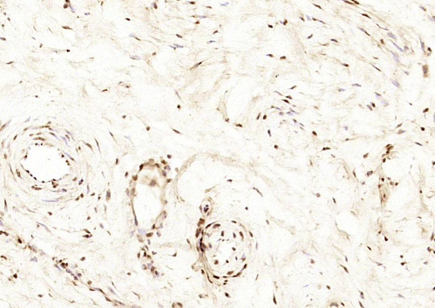
Paraformaldehyde-fixed, paraffin embedded (human cervical); Antigen retrieval by boiling in sodium citrate buffer (pH6.0) for 15min; Block endogenous peroxidase by 3% hydrogen peroxide for 20 minutes; Blocking buffer (normal goat serum) at 37°C for 30min; Antibody incubation with (Cytokeratin 4) Polyclonal Antibody, Unconjugated (bs-1006R) at 1:200 overnight at 4°C, followed by operating according to SP Kit(Rabbit) (sp-0023) instructionsand DAB staining.
-
Paraformaldehyde-fixed, paraffin embedded (human gastric); Antigen retrieval by boiling in sodium citrate buffer (pH6.0) for 15min; Block endogenous peroxidase by 3% hydrogen peroxide for 20 minutes; Blocking buffer (normal goat serum) at 37°C for 30min; Antibody incubation with (Cytokeratin 4) Polyclonal Antibody, Unconjugated (bs-1006R) at 1:200 overnight at 4°C, followed by operating according to SP Kit(Rabbit) (sp-0023) instructionsand DAB staining.
-
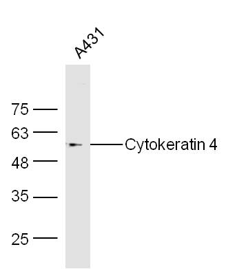
Sample:
A431(Human) Cell Lysate at 30 ug
Primary: Anti-Cytokeratin 4 (bs-1006R) at 1/300 dilution
Secondary: IRDye800CW Goat Anti-Rabbit IgG at 1/20000 dilution
Predicted band size: 57 kD
Observed band size: 57 kD
-
Tissue/cell: rat skin tissue; 4% Paraformaldehyde-fixed and paraffin-embedded;
Antigen retrieval: citrate buffer ( 0.01M, pH 6.0 ), Boiling bathing for 15min; Block endogenous peroxidase by 3% Hydrogen peroxide for 30min; Blocking buffer (normal goat serum,C-0005) at 37℃ for 20 min;
Incubation: Anti-CK4 Polyclonal Antibody, Unconjugated(bs-1006R) 1:200, overnight at 4°C, followed by conjugation to the secondary antibody(SP-0023) and DAB(C-0010) staining
-
Blank control:Hela.
Primary Antibody (green line): Rabbit Anti-Cytokeratin 4 antibody (bs-1006R)
Dilution: 1ug/Test;
Secondary Antibody : Goat anti-rabbit IgG-FITC
Dilution: 0.5ug/Test.
Protocol
The cells were fixed with 4% PFA (10min at room temperature)and then permeabilized with 90% ice-cold methanol for 20 min at -20℃.The cells were then incubated in 5%BSA to block non-specific protein-protein interactions for 30 min at room temperature .Cells stained with Primary Antibody for 30 min at room temperature. The secondary antibody used for 40 min at room temperature. Acquisition of 20,000 events was performed.
RRID:AB_10856840
产品名称:Rabbit Anti-Cytokeratin 4 antibody
别名: CK 4; CK4; CYK 4; CK-4; CYK4; Cytokeratin4; Cytokeratin-4; FLJ31692; K4; Keratin 4; Keratin-4; Keratin type II cytoskeletal 4; Keratin4; KRT 4; KRT4; K2C4_HUMAN; Keratin, type II cytoskeletal 4; K4; Type-II keratin Kb4.
中文名称:细胞角蛋白4抗体
英文名称:Rabbit Anti-Cytokeratin 4 antibody
抗体来源: Rabbit
克隆类型:多克隆
细胞定位:细胞核,细胞浆
性 状:Liquid
亚 型:IgG
纯化方法:affinity purified by Protein A
保存条件:Shipped at 4℃. Store at -20 °C for one year. Avoid repeated freeze/thaw cycles.
免 疫 原:KLH conjugated synthetic peptide derived from human CK4
抗原表位:256-360/594
SWISS:P19013
Gene ID :3851
Human Gene ID:3851
The protein encoded by this gene is a member of the keratin gene family. The type II cytokeratins consist of basic or neutral proteins which are arranged in pairs of heterotypic keratin chains coexpressed during differentiation of simple and stratified epithelial tissues. This type II cytokeratin is specifically expressed in differentiated layers of the mucosal and esophageal epithelia with family member KRT13. Mutations in these genes have been associated with White Sponge Nevus, characterized by oral, esophageal, and anal leukoplakia. The type II cytokeratins are clustered in a region of chromosome 12q12-q13. [provided by RefSeq, Jul 2008]
Subunit:Heterotetramer of two type I and two type II keratins. Keratin-4 is generally associated with keratin-13.
Tissue Specificity:Detected in the suprabasal layer of the stratified epithelium of the esophagus, exocervix, vagina, mouth and lingual mucosa, and in cells and cell clusters in the mucosa and serous gland ducts of the esophageal submucosa (at protein level). Expressed wide
DISEASE:Defects in KRT4 are a cause of white sponge nevus of cannon (WSN) [MIM:193900]. WSN is a rare autosomal dominant disorder which predominantly affects non-cornified stratified squamous epithelia. Clinically, it is characterized by the presence of soft, whi
Similarity:Belongs to the intermediate filament family.
Important Note:This product as supplied is intended for research use only, not for use in human, therapeutic or diagnostic applications.
 400-901-9800


 说明书
说明书 联系我们
联系我们 打印此页面
打印此页面 收藏
收藏