| Rabbit Anti-CD95/FAS antibody |
| 反应物种(预测) |
Pig |
| 产品应用(已验证) |
WB,FCM |
| 产品应用(可尝试) |
ELISA |
| 推荐稀释比例 |
WB=1:500-2000,Elisa=1:5000-10000,Flow Cyt=2μg/Test, |
| 研究领域 |
细胞生物,免疫学,细胞凋亡 |
| 标签 |
Array |
-
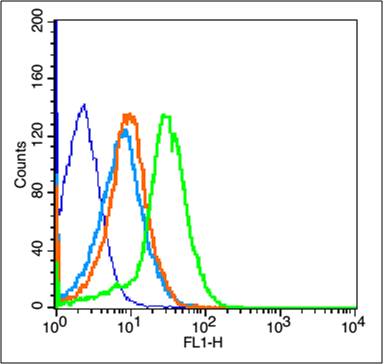
Blank control(blue):Mouse Kidney (fixed with 2% paraformaldehyde for 10 min at 37℃).
Primary Antibody:Rabbit Anti-CD95/FAS antibody (bs-6477R,Green); Dilution: 1μg in 100 μL 1X PBS containing 0.5% BSA;
Isotype Control Antibody: Rabbit IgG(orange) ,used under the same conditions;
Secondary Antibody: Goat anti-rabbit IgG-FITC(white blue), Dilution: 1:200 in 1 X PBS containing 0.5% BSA.
-
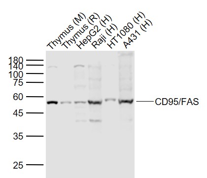
Sample:
Lane 1: Thymus (Mouse) Lysate at 40 ug
Lane 2: Thymus (Rat) Lysate at 40 ug
Lane 3: HepG2 (Human) Cell Lysate at 30 ug
Lane 4: Raji (Human) Cell Lysate at 30 ug
Lane 5: HT1080 (Human) Cell Lysate at 30 ug
Lane 6: A431 (Human) Cell Lysate at 30 ug
Primary: Anti- CD95/FAS (bs-6477R) at 1/1000 dilution
Secondary: IRDye800CW Goat Anti-Rabbit IgG at 1/20000 dilution
Predicted band size: 45/52 kD
Observed band size: 52 kD
-
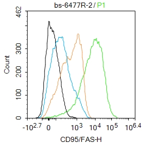
Blank control:Raji.
Primary Antibody (green line): Rabbit Anti-CD95/FAS antibody (bs-6477R)
Dilution: 2ug/Test;
Secondary Antibody : Goat anti-rabbit IgG-AF488
Dilution: 0.5ug/Test.
Protocol
The cells were incubated in 5%BSA to block non-specific protein-protein interactions for 30 min at room temperature .Cells stained with Primary Antibody for 30 min at room temperature. The secondary antibody used for 40 min at room temperature. Acquisition of 20,000 events was performed.
-
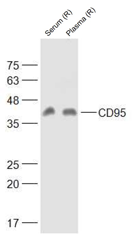
Sample:
serum (Rat) at 40 ug
plasma (Rat) at 40 ug
Primary: Anti-CD95 (bs-6477R) at 1/1000 dilution
Secondary: IRDye800CW Goat Anti-Rabbit IgG at 1/20000 dilution
Predicted band size: 34 kD
Observed band size: 42 kD
RRID:RRID
产品名称:Rabbit Anti-CD95/FAS antibody
别名: Apo-1; ALPS 1A; ALPS1A; APO 1; Apo 1 antigen; APO 1 cell surface antigen; Apo-1 antigen; APO1; Apo1 antigen; APO1 cell surface antigen; Apoptosis antigen 1; Apoptosis mediating surface antigen FAS; Apoptosis-mediating surface antigen FAS; APT 1; APT1; CD
中文名称:载脂蛋白1抗体
英文名称:Rabbit Anti-CD95/FAS antibody
抗体来源: Rabbit
克隆类型:多克隆
细胞定位:细胞膜,分泌型蛋白
性 状:Liquid
亚 型:IgG
纯化方法:affinity purified by Protein A
保存条件:Shipped at 4℃. Store at -20 °C for one year. Avoid repeated freeze/thaw cycles.
免 疫 原:KLH conjugated synthetic peptide derived from human FAS/Apo-1/CD95
抗原表位:81-170/335
抗原细胞定位:Extracellular
SWISS:P25445
Gene ID :355
Human Gene ID:355
The protein encoded by this gene is a member of the TNF-receptor superfamily. This receptor contains a death domain. It has been shown to play a central role in the physiological regulation of programmed cell death, and has been implicated in the pathogenesis of various malignancies and diseases of the immune system. The interaction of this receptor with its ligand allows the formation of a death-inducing signaling complex that includes Fas-associated death domain protein (FADD), caspase 8, and caspase 10. The autoproteolytic processing of the caspases in the complex triggers a downstream caspase cascade, and leads to apoptosis. This receptor has been also shown to activate NF-kappaB, MAPK3/ERK1, and MAPK8/JNK, and is found to be involved in transducing the proliferating signals in normal diploid fibroblast and T cells. Several alternatively spliced transcript variants have been described, some of which are candidates for nonsense-mediated mRNA decay (NMD). The isoforms lacking the transmembrane domain may negatively regulate the apoptosis mediated by the full length isoform. [provided by RefSeq, Mar 2011]
Function:Receptor for TNFSF6/FASLG. The adapter molecule FADD recruits caspase-8 to the activated receptor. The resulting death-inducing signaling complex (DISC) performs caspase-8 proteolytic activation which initiates the subsequent cascade of caspases (aspartat
Subunit:Binds DAXX. Interacts with HIPK3. Part of a complex containing HIPK3 and FADD. Binds RIPK1 and FAIM2. Interacts with BRE and FEM1B. Interacts with FADD.
Subcellular Location:Isoform 1: Cell membrane; Single-pass type I membrane protein. Isoform 2, 3, 4, 5, 6: Secreted.
Tissue Specificity:Isoform 1 and isoform 6 are expressed at equal levels in resting peripheral blood mononuclear cells. After activation there is an increase in isoform 1 and decrease in the levels of isoform 6.
Post-translational modifications:N- and O-glycosylated. O-glycosylated with core 1 or possibly core 8 glycans.
DISEASE:Defects in FAS are the cause of autoimmune lymphoproliferative syndrome type 1A (ALPS1A) [MIM:601859]; also known as Canale-Smith syndrome (CSS). ALPS is a childhood syndrome involving hemolytic anemia and thrombocytopenia with massive lymphadenopathy and
Similarity:Contains 1 death domain.
Contains 3 TNFR-Cys repeats.
Important Note:This product as supplied is intended for research use only, not for use in human, therapeutic or diagnostic applications.
 400-901-9800


 说明书
说明书 联系我们
联系我们 打印此页面
打印此页面 收藏
收藏